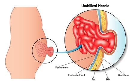
Umbilical hernia is a part of the intestine coming out of the umbilical opening in the abdominal muscles and is generally harmless. It is most commonly seen in infants. During the development of the baby in the womb, the cord that connects the mother to the baby is connected through a small opening in the baby's abdominal wall.
This opening should close itself shortly after birth. If not, the fatty tissue or part of the intestine leaves this opening and causes umbilical hernia. When the baby cries, it can be understood by protruding out of the navel…

How is it diagnosed and treated?
Umbilical hernia is a part of the intestine coming out of the umbilical opening in the abdominal muscles and is generally harmless. It is most commonly seen in infants. During the development of the baby in the womb, the cord that connects the mother to the baby is connected through a small opening in the baby's abdominal wall. This opening should close itself shortly after birth.
If not, the fatty tissue or part of the intestine leaves this opening and causes umbilical hernia. When the baby cries, it can be understood by protruding out of the navel. It usually recovers spontaneously until 1 or 2 years of age, but in some cases it may take longer to recover.
In adults, excessive pressure in the abdominal wall or a portion of the fatty tissue or intestine from the region where the abdominal muscles are weak causes hernia. The main causes are obesity, pushing and lifting heavy items, previous operations, and hernia history of the patient and her family.
Physical examination may reveal the presence of umbilical hernia. In some cases, imaging devices (such as ultrasound, CT) can be used to identify complications. Infants are expected to disappear spontaneously until the age of 4 years.
Surgical methods may be needed for the treatment of umbilical hernia occurring in children over the age of 4 or in adulthood. During surgery, a small incision in the umbilical pit is made and the hernia is returned to the abdominal cavity and the opening in the abdominal wall is closed by suturing.


